
MRI stands for Magnetic Resonance Imaging. An MRI scan uses radio waves strong magnetic fields to generate clear, detailed images of almost any part of the inside the body including:
- Bones
- Joints
- Brain
- Spinal Cord
- Breasts
- Heart
- Blood vessels
- Internal organs; womb, liver, kidney
An MRI scan is a way of efficiently diagnosing conditions, planning treatments and assessing how successful any previous treatment has been.
What happens during a MRI scan?
It is important to start by noting that an MRI scan is completely painless and extremely safe since the body is not being exposed to X-ray radiation.
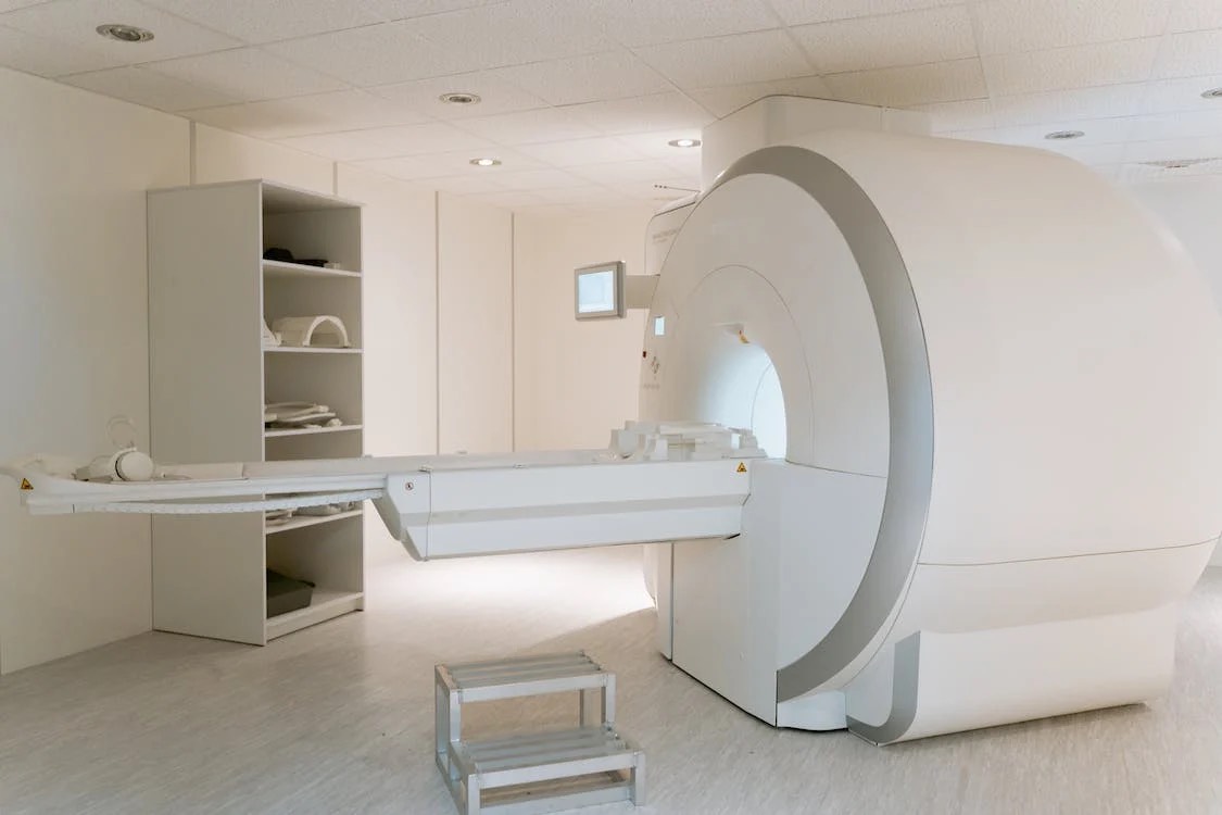
During the actual scan, you will be asked to lie flat on your back on a bed which is built for and moved into the scanner. You will either enter the scanner head first or feet first, depending on which part of the body is being scanned.
A ‘radiographer’ will be operating the scan, who is professionally trained in carrying out imaging investigations. The scanner is controlled via a computer in a separate room, since it cannot be near the magnetic field that the scanner generates.
You will be able to communicate through an intercom during the scan, and the radiographer will be able to see you on a monitor. Having this knowledge makes people feel a lot better about undergoing a scan, as many feel nervous that they will feel overwhelmingly claustrophobic. The ability to communicate and know that you can be seen on a TV screen should put your mind at ease.
Since there is certain to be a loud tapping noise at some points during the scan, you will be given earplugs or headphones. The noise is just the electric current in the scanner’s coils being turned on and off – so there is nothing to worry about there!
MRI scans require you to keep as still as possible for the entire scan which can last between 15 – 90 minutes. The length of the scan depends on the size of the area that is being looked at.
How does an MRI scan work?
According to the NHS, the majority of the human body is made up of water molecules, consisting of oxygen and hydrogen atoms. At the centre of the hydrogen atom is a much smaller particle, known as a proton. Like tiny magnets, protons are highly sensitive to magnetic fields. When exposed to powerful magnets, the protons line up in the same direction.
Radio waves are sent to certain areas of the body in short bursts, which knock the protons out of alignment. The protons realign when the radio waves are turned off. Radio signals are sent out, to then be picked up by receivers. These signals provide the information needed about the protons of an exact location of the body. The signals from these protons are combined to create the detailed image of the inside of the body, just like pixels on a computer creates detailed images on the screen.
MRI scans have been found to be one of the safest, current medical procedures as no evidence has be found to suggest that the radio waves or magnetic fields pose harm to the body. However, it is unsafe and therefore not offered to people who have certain types of implants fitted, for example a pacemaker.